Category:Signal transduction
Jump to navigation
Jump to search

| Category Signal transduction on sister projects: | ||||||||||||||||||||
|---|---|---|---|---|---|---|---|---|---|---|---|---|---|---|---|---|---|---|---|---|
en:
|
||||||||||||||||||||
Subcategories
This category has the following 22 subcategories, out of 60 total.
(previous page) (next page)P
- Paxillin (27 F)
- Protein kinase C signaling (16 F)
- Protein kinase signaling cascade (53 F)
R
- Response elements (12 F)
S
- Second messenger systems (10 F)
- Secretory pathway (102 F)
- STAT signaling family (5 F)
- Stress signaling cascade (11 F)
- Synaptic active zone (2 F)
T
- Tor signaling (4 F)
- Transcriptional signaling (8 F)
Media in category "Signal transduction"
The following 200 files are in this category, out of 1,548 total.
(previous page) (next page)-
-
-
-
-
 P53PPINetworkFromIntact.png 1,280 × 1,024; 1.73 MB
-
 Panel-Call-Indicator-Code.png 2,460 × 1,404; 2.89 MB
-
-
 PBB Protein STK10 image.jpg 500 × 500; 53 KB
-
-
-
-
-
-
-
-
-
-
-
-
-
-
Persistent-Cellular-Motion-Control-and-Trapping-Using-Mechanotactic-Signaling-pone.0105406.s008.ogv 20 s, 900 × 760; 2.38 MB
-
Persistent-Cellular-Motion-Control-and-Trapping-Using-Mechanotactic-Signaling-pone.0105406.s009.ogv 24 s, 900 × 760; 3.59 MB
-
Persistent-Cellular-Motion-Control-and-Trapping-Using-Mechanotactic-Signaling-pone.0105406.s010.ogv 32 s, 900 × 760; 3.54 MB
-
-
-
-
-
-
-
-
-
-
-
 PhK 1QL6 gamma small.png 346 × 402; 61 KB
-
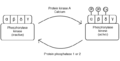 PhK diagram.png 645 × 327; 9 KB
-
-
 Photorezeptor.png 352 × 395; 7 KB
-
 PI3K inhibitors overview Mishra2021.jpg 775 × 773; 130 KB
-
PI3Ks-Maintain-the-Structural-Integrity-of-T-Tubules-in-Cardiac-Myocytes-pone.0024404.s004.ogv 5.0 s, 656 × 752; 2.22 MB
-
PI3Ks-Maintain-the-Structural-Integrity-of-T-Tubules-in-Cardiac-Myocytes-pone.0024404.s005.ogv 5.0 s, 656 × 752; 2.34 MB
-
PINK1-Mediated-Phosphorylation-of-Parkin-Boosts-Parkin-Activity-in-Drosophila-pgen.1004391.s006.ogv 32 s, 640 × 480; 1.08 MB
-
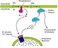
 PIP2 cleavage by PLC to release IP3 and DAG tr.png 1,171 × 977; 188 KB
-
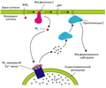 PIP2 cleavage by PLC to release IP3 and DAG uk.png 1,171 × 977; 186 KB
-
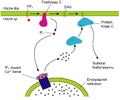
 PIP2 cleavage to IP3 and DAG.jpg 1,171 × 977; 204 KB
-
 Piskacek p53a.jpg 7,568 × 2,528; 1.8 MB
-
 Piskacek p53b.jpg 5,344 × 2,000; 1.64 MB
-
-
-
Plant-Kinesin-Like-Calmodulin-Binding-Protein-Employs-Its-Regulatory-Domain-for-Dimerization-pone.0066669.s002.ogv 8.7 s, 1,392 × 1,040; 11.94 MB
-
Plant-Kinesin-Like-Calmodulin-Binding-Protein-Employs-Its-Regulatory-Domain-for-Dimerization-pone.0066669.s003.ogv 8.7 s, 1,392 × 1,040; 8.8 MB
-
-
-
Plasticity-in-the-Macromolecular-Scale-Causal-Networks-of-Cell-Migration-pone.0090593.s007.ogv 8.2 s, 3,072 × 1,024; 3.89 MB
-
Plasticity-in-the-Macromolecular-Scale-Causal-Networks-of-Cell-Migration-pone.0090593.s008.ogv 9.7 s, 3,072 × 1,024; 6.51 MB
-
Plasticity-in-the-Macromolecular-Scale-Causal-Networks-of-Cell-Migration-pone.0090593.s009.ogv 8.1 s, 2,150 × 716; 964 KB
-
PLCγ-activated-signalling-is-essential-for-TrkB-mediated-sensory-neuron-structural-plasticity-1471-213X-10-103-S1.ogv 2 min 0 s, 320 × 240; 1.37 MB
-
PLCγ-activated-signalling-is-essential-for-TrkB-mediated-sensory-neuron-structural-plasticity-1471-213X-10-103-S2.ogv 2 min 0 s, 320 × 240; 1.29 MB
-
-
-
-
-
-
-
-
-
-
-
-
Possible-Involvement-of-Cone-Opsins-in-Distinct-Photoresponses-of-Intrinsically-Photosensitive-pone.0070342.s001.ogv 16 s, 1,440 × 1,080; 3.53 MB
-
-
-
 PPAR transrepression.svg 1,655 × 967; 31 KB
-
Primary-Neuron-Culture-for-Nerve-Growth-and-Axon-Guidance-Studies-in-Zebrafish-(Danio-rerio)-pone.0057539.s003.ogv 28 s, 1,388 × 1,040; 12.43 MB
-
-
ProBDNF-Collapses-Neurite-Outgrowth-of-Primary-Neurons-by-Activating-RhoA-pone.0035883.s001.ogv 7.1 s, 800 × 600; 1.41 MB
-
ProBDNF-Collapses-Neurite-Outgrowth-of-Primary-Neurons-by-Activating-RhoA-pone.0035883.s002.ogv 12 s, 800 × 600; 2.5 MB
-
-
-
-
-
-
-
-
-
-
-
-
-
-
-
-
-
-
-
-
-
-
Protein-receptor-independent-plasma-membrane-remodeling-by-HAMLET-a-tumoricidal-protein-lipid-srep16432-s5.ogv 3 min 8 s, 512 × 512; 2.65 MB
-
Protein-receptor-independent-plasma-membrane-remodeling-by-HAMLET-a-tumoricidal-protein-lipid-srep16432-s6.ogv 1 min 6 s, 512 × 512; 1.42 MB
-
Protein-receptor-independent-plasma-membrane-remodeling-by-HAMLET-a-tumoricidal-protein-lipid-srep16432-s7.ogv 1 min 6 s, 512 × 512; 971 KB
-
-
-
 Psy161ST redrawn.svg 951 × 175; 40 KB
-
Ptenb-Mediates-Gastrulation-Cell-Movements-via-Cdc42AKT1-in-Zebrafish-pone.0018702.s002.ogv 14 s, 786 × 626; 5.03 MB
-
Ptenb-Mediates-Gastrulation-Cell-Movements-via-Cdc42AKT1-in-Zebrafish-pone.0018702.s003.ogv 17 s, 786 × 626; 5.61 MB
-
Ptenb-Mediates-Gastrulation-Cell-Movements-via-Cdc42AKT1-in-Zebrafish-pone.0018702.s005.ogv 8.7 s, 868 × 716; 913 KB
-
Ptenb-Mediates-Gastrulation-Cell-Movements-via-Cdc42AKT1-in-Zebrafish-pone.0018702.s006.ogv 15 s, 512 × 512; 102 KB
-
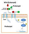 QseC Signaltransduktion.jpg 850 × 980; 262 KB
-
-
-
-
-
-
-
-
-
-
-
-
-
-
-
-
-
-
-
-
-
-
-
-
-
-
-
-
-
Reactive-Oxygen-Species-Regulate-Protrusion-Efficiency-by-Controlling-Actin-Dynamics-pone.0041342.s008.ogv 6.1 s, 526 × 1,110; 6.08 MB
-
-
-
Reactive-Oxygen-Species-Regulate-Protrusion-Efficiency-by-Controlling-Actin-Dynamics-pone.0041342.s011.ogv 3.0 s, 510 × 1,242; 3.64 MB
-
-
-
-
-
-
-
-
-
-
 Regulation of mTOR signaling through primary cilia.png 2,155 × 2,571; 527 KB
-
-
-
-
-
-
-
-
-
-
Regulation-of-Embryonic-Cell-Adhesion-by-the-Prion-Protein-pbio.1000055.sv001.ogv 8.0 s, 299 × 299; 1.19 MB
-
Regulation-of-Embryonic-Cell-Adhesion-by-the-Prion-Protein-pbio.1000055.sv002.ogv 8.0 s, 356 × 357; 1.66 MB
-
Regulation-of-Embryonic-Cell-Adhesion-by-the-Prion-Protein-pbio.1000055.sv003.ogv 12 s, 158 × 155; 88 KB
-
Regulation-of-Embryonic-Cell-Adhesion-by-the-Prion-Protein-pbio.1000055.sv004.ogv 12 s, 158 × 156; 92 KB
-
Regulation-of-Embryonic-Cell-Adhesion-by-the-Prion-Protein-pbio.1000055.sv005.ogv 2.1 s, 1,024 × 1,024; 1.68 MB
-
Regulation-of-Embryonic-Cell-Adhesion-by-the-Prion-Protein-pbio.1000055.sv006.ogv 12 s, 523 × 512; 340 KB
-
-
-
-
-
-
-
-
-
-
-
-
-
-
-
-
-
-
-
-
-
-
-
-
 RegulationOfAMPARTrafficking-ukr.jpg 1,162 × 908; 239 KB
-
 RegulationOfAMPARTrafficking.jpg 1,162 × 908; 104 KB
-
-
-
-
-
Repressor-Dimerization-in-the-Zebrafish-Somitogenesis-Clock-pcbi.0030032.sv001.ogv 20 s, 640 × 480; 3.15 MB
-
Repressor-Dimerization-in-the-Zebrafish-Somitogenesis-Clock-pcbi.0030032.sv002.ogv 13 s, 640 × 480; 2.89 MB
-
Repressor-Dimerization-in-the-Zebrafish-Somitogenesis-Clock-pcbi.0030032.sv003.ogv 13 s, 640 × 480; 2.81 MB
-
Repressor-Dimerization-in-the-Zebrafish-Somitogenesis-Clock-pcbi.0030032.sv004.ogv 6.7 s, 640 × 480; 1.12 MB
-
Repressor-Dimerization-in-the-Zebrafish-Somitogenesis-Clock-pcbi.0030032.sv005.ogv 6.7 s, 640 × 480; 1.14 MB


