Neurilemma: Difference between revisions
Hydrargyrum (talk | contribs) t |
Remove vandalism written by Blay Ambrose |
||
| (33 intermediate revisions by 17 users not shown) | |||
| Line 1: | Line 1: | ||
{{Short description|Layer present on Schwann cells of PNS neurons}}{{Infobox microanatomy |
|||
{{Infobox anatomy |
|||
| Name = |
| Name = Neurilemma |
||
| Latin = |
| Latin = |
||
| GraySubject = 183 |
|||
| GrayPage = 727 |
|||
| Image = Gray631.png |
| Image = Gray631.png |
||
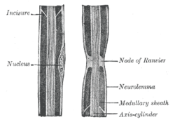
| Caption = Diagram of longitudinal sections of medullated nerve fibers. |
| Caption = Diagram of longitudinal sections of medullated nerve fibers. |
||
| Width = |
| Width = |
||
| Image2 = |
| Image2 = Myelin sheath (1).svg| |
||
| Caption2 = Transverse sections of medullated nerve fibers. |
|||
| Caption2 = Cross section of an axon.<br /> |
|||
| ImageMap = |
|||
1. Axon<br /> |
|||
| MapCaption = |
|||
2. Nucleus of Schwann cell<br /> |
|||
| Precursor = |
|||
3. [[Schwann cell]]<br /> |
|||
| System = |
|||
4. [[Myelin sheath]]<br /> |
|||
| Artery = |
|||
5. '''Neurilemma''' |
|||
| Vein = |
|||
| Location = [[Schwann cell]] |
|||
| Nerve = |
|||
| System = [[Peripheral nervous system]] |
|||
| Lymph = |
|||
| MeshName = A08.561.600 |
|||
| MeshNumber = Neurilemma |
|||
| Code = {{TerminologiaHistologica|2|00|06.1.00002| }} |
|||
| Dorlands = six/000071648 |
|||
| DorlandsID = Neurilemma |
|||
}} |
}} |
||
''' |
'''Neurilemma''' (also known as '''neurolemma''', '''sheath of Schwann''', or '''Schwann's sheath''')<ref name="Dorlands">{{cite book|last1=Albert|first1=Daniel|title=Dorland's illustrated medical dictionary.|date=2012|publisher=Saunders/Elsevier|location=Philadelphia, PA|isbn=9781416062578|pages=1262–1263|edition= 32nd}}</ref> is the outermost [[cell nucleus|nucleated]] [[cytoplasm]]ic layer of [[Schwann cell]]s (also called neurilemmocytes) that surrounds the [[axon]] of the [[neuron]]. It forms the outermost layer of the [[nerve fiber]] in the [[peripheral nervous system]].<ref name="Marieb" >{{cite book |author1=Elaine N. Marieb |author2=Katja Hoehn | title = Human Anatomy & Physiology (7th Ed.) |url=https://archive.org/details/humananatomyphys00mari_4 |url-access=registration | publisher = Pearson | pages = [https://archive.org/details/humananatomyphys00mari_4/page/394 394–5] | year = 2007 | isbn = 978-0-8053-5909-1}}</ref> |
||
The |
The neurilemma is underlain by the [[myelin |myelin sheath]] (also known as the medullary sheath). In the [[central nervous system]], axons are myelinated by [[oligodendrocytes]], thus lack neurilemma. The myelin sheaths of oligodendrocytes do not have neurilemma because excess [[cytoplasm]] is directed centrally toward the oligodendrocyte cell body. |
||
Neurilemma serves a protective function for peripheral nerve fibers. Damaged nerve fibers may regenerate if the [[Soma (biology)|cell body]] is not damaged and the neurilemma remains intact. The neurilemma forms a regeneration tube through which the growing axon re-establishes its original connection. |
|||
[[Neurilemoma]] is a tumor of the neurilemma.<ref name=Dorlands/> |
|||
==References== |
==References== |
||
{{ |
{{Reflist|1}} |
||
==External links== |
==External links== |
||
* [http://www.dmacc.edu/instructors/periphxs.htm Histology at dmacc.edu] |
* [https://web.archive.org/web/20070212061042/http://www.dmacc.edu/instructors/periphxs.htm Histology at dmacc.edu] |
||
| ⚫ | |||
[[Category:Neuroanatomy]] |
|||
| ⚫ | |||
{{Nervous tissue}} |
{{Nervous tissue}} |
||
{{Authority control}} |
|||
| ⚫ | |||
| ⚫ | |||
Latest revision as of 20:34, 24 September 2022
| Neurilemma | |
|---|---|
 Diagram of longitudinal sections of medullated nerve fibers. | |
 Cross section of an axon. 1. Axon | |
| Details | |
| System | Peripheral nervous system |
| Standort | Schwann cell |
| Identifiers | |
| MeSH | D009441 |
| TH | H2.00.06.1.00002 |
| Anatomical terms of microanatomy | |
Neurilemma (also known as neurolemma, sheath of Schwann, or Schwann's sheath)[1] is the outermost nucleated cytoplasmic layer of Schwann cells (also called neurilemmocytes) that surrounds the axon of the neuron. It forms the outermost layer of the nerve fiber in the peripheral nervous system.[2]
The neurilemma is underlain by the myelin sheath (also known as the medullary sheath). In the central nervous system, axons are myelinated by oligodendrocytes, thus lack neurilemma. The myelin sheaths of oligodendrocytes do not have neurilemma because excess cytoplasm is directed centrally toward the oligodendrocyte cell body.
Neurilemma serves a protective function for peripheral nerve fibers. Damaged nerve fibers may regenerate if the cell body is not damaged and the neurilemma remains intact. The neurilemma forms a regeneration tube through which the growing axon re-establishes its original connection.
Neurilemoma is a tumor of the neurilemma.[1]
References
[edit]- ^ a b Albert, Daniel (2012). Dorland's illustrated medical dictionary (32nd ed.). Philadelphia, PA: Saunders/Elsevier. pp. 1262–1263. ISBN 9781416062578.
- ^ Elaine N. Marieb; Katja Hoehn (2007). Human Anatomy & Physiology (7th Ed.). Pearson. pp. 394–5. ISBN 978-0-8053-5909-1.
External links
[edit]
