
Collateral fissure: Difference between revisions
Content deleted Content added
m cleanup (wikitables, html markup, layout, etc.) |
SimLibrarian (talk | contribs) mNo edit summary Tags: Mobile edit Mobile app edit iOS app edit |
||
| (9 intermediate revisions by 7 users not shown) | |||
| Line 1: | Line 1: | ||
{{Short description|Brain structure}} |
|||
{{Infobox brain |
{{Infobox brain |
||
| Name = Collateral fissure |
| Name = Collateral fissure |
||
| Latin = sulcus collateralis, fissura collateralis |
| Latin = sulcus collateralis, fissura collateralis |
||
| ⚫ | |||
| GraySubject = 189 |
|||
| ⚫ | |||
| GrayPage = 820 |
|||
| |
| Image2 = OccCaptsMedial.png |
||
| Caption2 = Medial surface of right cerebral hemisphere. Collateral sulcus divides [[Limbic lobe|limbic]] (purple) and [[temporal lobe]] (green). |
|||
| ⚫ | |||
| ⚫ | |||
| ⚫ | |||
| IsPartOf = |
| IsPartOf = |
||
| Components = |
| Components = |
||
| Artery = |
| Artery = |
||
| Vein = |
| Vein = |
||
| BrainInfoType = hier |
|||
| BrainInfoNumber = 28 |
|||
| MeshName = |
|||
| MeshNumber = |
|||
| DorlandsPre = f_08 |
|||
| DorlandsSuf = 12365914 |
|||
}} |
}} |
||
| Line 24: | Line 17: | ||
Behind, it lies below and lateral to the [[calcarine fissure]], from which it is separated by the [[lingual gyrus]]; in front, it is situated between the [[parahippocampal gyrus]] and the anterior part of the [[fusiform gyrus]]. |
Behind, it lies below and lateral to the [[calcarine fissure]], from which it is separated by the [[lingual gyrus]]; in front, it is situated between the [[parahippocampal gyrus]] and the anterior part of the [[fusiform gyrus]]. |
||
== Additional images == |
|||
| ⚫ | |||
<gallery> |
|||
| ⚫ | |||
File:Hippocampal Limbic Connections Functions - Sanjoy Sanyal (Cropped from 5m28s to 6m30s) Collateral sulcus.webm|Human brain dissection video (62 sec). Demonstrating location of collateral sulcus. |
|||
</gallery> |
|||
| ⚫ | |||
== References == |
|||
{{Gray's}} |
{{Gray's}} |
||
{{Telencephalon}} |
{{Telencephalon}} |
||
{{Portal bar|Anatomy}} |
|||
{{Authority control}} |
|||
[[Category:Cerebrum]] |
[[Category:Cerebrum]] |
||
[[Category:Sulci (neuroanatomy)]] |
[[Category:Sulci (neuroanatomy)]] |
||
[[Category:Articles containing video clips]] |
|||
{{ |
{{neuroanatomy-stub}} |
||
Latest revision as of 17:48, 21 November 2023
| Collateral fissure | |
|---|---|
 Medial surface of left cerebral hemisphere. (Collateral fissure labeled at bottom left.) | |
 Medial surface of right cerebral hemisphere. Collateral sulcus divides limbic (purple) and temporal lobe (green). | |
| Details | |
| Identifiers | |
| Latin | sulcus collateralis, fissura collateralis |
| NeuroNames | 47 |
| TA98 | A14.1.09.206 |
| TA2 | 5442 |
| FMA | 83751 |
| Anatomical terms of neuroanatomy | |
The collateral fissure (or sulcus) is on the tentorial surface of the hemisphere and extends from near the occipital pole to within a short distance of the temporal pole.
Behind, it lies below and lateral to the calcarine fissure, from which it is separated by the lingual gyrus; in front, it is situated between the parahippocampal gyrus and the anterior part of the fusiform gyrus.
Additional images
[edit]-
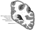 Coronal section through posterior cornua of lateral ventricle. (Collateral fissure labeled at bottom center.)
 -
Human brain dissection video (62 sec). Demonstrating location of collateral sulcus.

Wikimedia Commons has media related to Collateral sulcus.
References
[edit]![]() This article incorporates text in the public domain from page 820 of the 20th edition of Gray's Anatomy (1918)
This article incorporates text in the public domain from page 820 of the 20th edition of Gray's Anatomy (1918)
This neuroanatomy article is a stub. You can help Wikipedia by expanding it. |