
Microcalcification: Difference between revisions
m +cat, stub |
m fixed lint errors – file options; thumbnails shouldn't be used in galleries |
||
| (31 intermediate revisions by 20 users not shown) | |||
| Line 1: | Line 1: | ||
{{Short description|Calcium deposits in the breast}} |
|||
'''Microcalcifications''' are tiny specks of [[mineral deposit]]s ([[calcium]]), that can be scattered throughout the [[mammary gland]], or occur in clusters. When found on a [[mammogram]], an operative will then decide whether the specks are of concern - usually, this is not the case. Commonly, they simply indicate the presence of tiny benign [[cyst]]s, but can signify the presence of early [[breast cancer]]; for this reason, it is important to attend regular screening sessions, as recommended by your health service. |
|||
[[File:Mammogram microcalcifications in carcinoma in situ, CC, details.png|thumb|upright=1.3|[[Mammogram]] microcalcifications in [[ductal carcinoma in situ]]]] |
|||
'''Microcalcifications''' are tiny [[mineral deposit|deposits]] of [[calcium]] salts that are too small to be felt but can be detected by [[medical imaging|imaging]].<ref>{{cite web |title=NCI Dictionary of Cancer Terms |url=https://www.cancer.gov/publications/dictionaries/cancer-terms/def/microcalcification |website=National Cancer Institute |accessdate=23 April 2019 |language=en |date=2 February 2011}}</ref> |
|||
They are caused by a number of reasons: |
|||
They can be scattered throughout the [[mammary gland]], or occur in clusters. |
|||
Aging - the majority of diagnoses are made in women over 50 |
|||
Microcalcifications can be an early sign of [[breast cancer]]. Based on morphology, it is possible to classify by radiography how likely microcalcifications are to indicate cancer. |
|||
Benign causes |
|||
<ref>{{cite journal |last1=Nalawade |first1=Yojana V |title=Evaluation of breast calcifications |journal=The Indian Journal of Radiology & Imaging |date=November 2009 |volume=19 |issue=4 |pages=282–286 |doi=10.4103/0971-3026.57208 |pmid=19881103 |issn=0971-3026|pmc=2797739 |doi-access=free }}</ref> |
|||
Genetic - involving the [[BRCA1 gene]] and [[BRCA2 gene]]s |
|||
==In breast== |
|||
Depending on what pattern the microcalcifications present determines the future course of the action, whether it be further investigatory techniques (as part of the triple assessment), or more regular screening. |
|||
Microcalcifications in the [[breast]] are made up of [[calcium phosphate]] or [[calcium oxalate]]. When consisting of calcium phosphate, they are usually [[dystrophic calcifications]] (occurring in degenerated or necrotic tissue).<ref name="pmid35251632">{{cite journal| author=Logullo AF, Prigenzi KCK, Nimir CCBA, Franco AFV, Campos MSDA| title=Breast microcalcifications: Past, present and future (Review). | journal=Mol Clin Oncol | year= 2022 | volume= 16 | issue= 4 | pages= 81 | pmid=35251632 | doi=10.3892/mco.2022.2514 | pmc=8892454 | url=https://www.ncbi.nlm.nih.gov/entrez/eutils/elink.fcgi?dbfrom=pubmed&tool=sumsearch.org/cite&retmode=ref&cmd=prlinks&id=35251632 }} </ref> Yet, the mechanism of their formation is not fully known.<ref name="wilkinson">{{cite journal |last1=Wilkinson |first1=Louise |last2=Thomas |first2=Val |last3=Sharma |first3=Nisha |title=Microcalcification on mammography: approaches to interpretation and biopsy |journal=The British Journal of Radiology |volume=90 |issue=1069 |pages=20160594 |doi=10.1259/bjr.20160594 |pmid=27648482 |pmc=5605030 |issn=0007-1285|year=2016 }}</ref> |
|||
Calcium oxalate crystals in the breast may be seen on [[mammography]] and are usually benign, but can be associated with [[lobular carcinoma in situ]].<ref>{{cite web|url=https://www.pathologyoutlines.com/topic/breastcalcification.html|title=Microcalcifications|author=Hind Warzecha, M.D.|website=Pathology Outlines}} Last author update: 1 June 2010</ref> |
|||
| ⚫ | |||
Microcalcification was first described in 1913 by surgeon [[Albert Salomon (surgeon)|Albert Salomon]].<ref name=wilkinson/> |
|||
| ⚫ | |||
<gallery> |
|||
File:Histopathology of microcalcifications in non-neoplastic breast.jpg|Calcium phosphate microcalcifications in non-neoplastic breast tissue. |
|||
File:Histopathology of dystrophic microcalcifications in ductal carcinoma in situ.jpg|Histopathology of dystrophic calcium phosphate microcalcifications in [[ductal carcinoma in situ]] (DCIS) of the breast, H&E stain. |
|||
File:Histopathology of a breast cyst with calcium oxalate crystals, annotated.jpg|Histopathology of calcium oxalate crystals in a benign breast cyst, H&E stain. |
|||
</gallery> |
|||
In contrast to an artifact of crowded cells, the DCIS calcification pictured above characteristically extends outside the focal plane, as the background DCIS is blurred in this focus. |
|||
== References == |
|||
{{reflist}} |
|||
| ⚫ | |||
| ⚫ | |||
Latest revision as of 22:28, 8 December 2023

Microcalcifications are tiny deposits of calcium salts that are too small to be felt but can be detected by imaging.[1]
They can be scattered throughout the mammary gland, or occur in clusters. Microcalcifications can be an early sign of breast cancer. Based on morphology, it is possible to classify by radiography how likely microcalcifications are to indicate cancer. [2]
In breast
[edit]Microcalcifications in the breast are made up of calcium phosphate or calcium oxalate. When consisting of calcium phosphate, they are usually dystrophic calcifications (occurring in degenerated or necrotic tissue).[3] Yet, the mechanism of their formation is not fully known.[4]
Calcium oxalate crystals in the breast may be seen on mammography and are usually benign, but can be associated with lobular carcinoma in situ.[5]
Microcalcification was first described in 1913 by surgeon Albert Salomon.[4]
-
 Calcium phosphate microcalcifications in non-neoplastic breast tissue.
 -
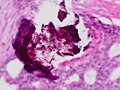 Histopathology of dystrophic calcium phosphate microcalcifications in ductal carcinoma in situ (DCIS) of the breast, H&E stain.
 -
 Histopathology of calcium oxalate crystals in a benign breast cyst, H&E stain.


In contrast to an artifact of crowded cells, the DCIS calcification pictured above characteristically extends outside the focal plane, as the background DCIS is blurred in this focus.
References
[edit]- ^ "NCI Dictionary of Cancer Terms". National Cancer Institute. 2 February 2011. Retrieved 23 April 2019.
- ^ Nalawade, Yojana V (November 2009). "Evaluation of breast calcifications". The Indian Journal of Radiology & Imaging. 19 (4): 282–286. doi:10.4103/0971-3026.57208. ISSN 0971-3026. PMC 2797739. PMID 19881103.
- ^ Logullo AF, Prigenzi KCK, Nimir CCBA, Franco AFV, Campos MSDA (2022). "Breast microcalcifications: Past, present and future (Review)". Mol Clin Oncol. 16 (4): 81. doi:10.3892/mco.2022.2514. PMC 8892454. PMID 35251632.
{{cite journal}}: CS1 maint: multiple names: authors list (link) - ^ a b Wilkinson, Louise; Thomas, Val; Sharma, Nisha (2016). "Microcalcification on mammography: approaches to interpretation and biopsy". The British Journal of Radiology. 90 (1069): 20160594. doi:10.1259/bjr.20160594. ISSN 0007-1285. PMC 5605030. PMID 27648482.
- ^ Hind Warzecha, M.D. "Microcalcifications". Pathology Outlines. Last author update: 1 June 2010
This medical sign article is a stub. You can help Wikipedia by expanding it. |