
Fascia: Difference between revisions
mNo edit summary |
added Fascial Dynamics section |
||
| Line 34: | Line 34: | ||
Image:Gray1125.png|The [[peritoneum]] and [[renal fascia]] |
Image:Gray1125.png|The [[peritoneum]] and [[renal fascia]] |
||
</gallery> |
</gallery> |
||
==Fascial Dynamics== |
|||
Fascia is a highly adaptable tissue. Due to its [[elastic]] property, superficial fascia can stretch to accomodate the deposition of adipose that accompanies both ordinary and [[prenatal]] weight gain. After [[pregnancy]] and weight loss, the superficial fascia slowly reverts to its original level of tension. |
|||
Deep fascia is less extensible than superficial fascia. It is essentially [[avascular]] <ref> {{cite book | last = Rolf | first = Ida P. | title = Rolfing | publisher = Healing Arts Press | date = 1989 | location = Rochester, VT | pages = 38 | id = ISBN 0-89281-335-0}} </ref>, but is richly [[innervated]] with [[sensory receptor]]s that report the presence of pain ([[nociceptors]]); change in movement ([[proprioceptors]]); change in pressure and vibration ([[mechanoreceptors]]); change in the chemical milieu ([[chemoreceptors]]); and fluctuation in temperature ([[thermoreceptors]]). <ref> {{cite book | last = Chaitow | first = Leon | title = Soft Tissue Manipulation | publisher = Healing Arts Press | date = 1988 | location = Rochester, VT | pages = 26-28 | | id = ISBN 0-89281-276-1 }} </ref>, <ref> {{cite journal | last = Schleip | first = R. | title = Fascial plasticity – a new neurobiological explanation: Part 1| journal = Journal of Bodywork and Movement Therapies | volume = 7 | issue = 1 | pages = 15-19 | publisher = Elsevier | date = 2003 |}} </ref> Deep fascia is able to respond to sensory input by contracting; by relaxing; or by adding, reducing, or changing its composition through the process of fascial remodeling. <ref> {{cite book | last = Myers | first = Thomas W. | title = Anatomy Trains | publisher = Churchill Livingstone | date = 2002 | location = London, UK | pages = 15 | id = ISBN 0-443-06351-6}} </ref> |
|||
Deep fascia can contract. What happens during the [[fight-or-flight response]] is an example of rapid fascial contraction . In response to a real or imagined threat to the organism, the body responds with a temporary increase in the stiffness of the fascia. Bolstered with tensioned fascia, people are able to perform extraordinary feats of strength and speed under emergency conditions. <ref> {{cite journal | last = Schleip | first = R. | authorlink = | coauthors = Klingler W.; Lehmann-Horn, F. | title = Active fascial contractility: Fascia may be able to contract in a smooth muscle-like manner and thereby influence musculoskeletal dynamics | journal = Medical Hypotheses | volume = 65 | pages = 274 | publisher = Elsevier | date = 2005 |}} </ref> How fascia contracts is still not well understood, but appears to involve the activity of [[myofibroblasts]]. Myofibroblasts are fascial cells that are created as a response to mechanical stress. In a two step process, [[fibroblasts]] differentiate into proto-myofibroblasts that with continued mechanical stress, become differentiated myofibroblasts. <ref> {{cite journal | last = Tomasek | first = J. | authorlink = | coauthors = Gabbiani, G.; Hinz, B.; Chaponnier, C.; Brown, R. | title = Myofibroblasts and Mechanoregulation of Connective Tissue Remodelling | journal = Molecular Cell Biology | volume = 3 | pages = 350-352 | publisher = Nature Publishing Group | date = 2002 |}} </ref> Fibroblasts cannot contract, but myofibroblasts are able to contract in a [[smooth muscle]]-like manner. <ref> {{cite journal | last = Schleip | first = R. | authorlink = | coauthors = Klingler, W.; Lehmann-Horn, F. | title = Active fascial contractility: Fascia may be able to contract in a smooth muscle-like manner and thereby influence musculoskeletal dynamics | journal = Medical Hypotheses | volume = 65 | pages = 273-277 | publisher = Elsevier | date = 2005 |}} </ref> If fascial contraction persists, fibroblasts secrete more collagen into the [[extracellular matrix]], remodeling the fascia to be thicker and less extensible. |
|||
The deep fascia can also relax. By monitoring changes in muscular tension, joint position, rate of movement, pressure, and vibration, mechanoreceptors in the deep fascia are capable of initiating relaxation. Deep fascia can relax rapidly in response to sudden muscular overload or rapid movements. [[Golgi tendon organs]] operate as a feedback mechanism by causing myofascial relaxation before muscle force becomes so great that tendons might be torn. [[Pacinian corpuscles]] monitor the rate of [[acceleration]] of movement and will intiate a sudden relaxatory response if movement happens too fast. <ref> {{cite book | last = Chaitow | first = Leon | title = Soft Tissue Manipulation | publisher = Healing Arts Press | date = 1988 | location = Rochester, VT | pages = 26-27 | | id = ISBN 0-89281-276-1 }} </ref> Deep fascia can also relax slowly as some mechanoreceptors are designed to report changes over a longer period of time. Unlike the Golgi tendon organs, Golgi receptors report joint position independent of muscle contraction. This helps the body to know where the bones are at any given moment. [[Ruffini endings]] respond to regular stretching and to slow sustained pressure. In addition to initiating fascial relaxation, they also have been shown to inhibit sympathetic activity by slowing down heart rate and respiration. <ref> {{cite journal | last = Schleip | first = R. | title = Fascial plasticity – a new neurobiological explanation: Part 1 | journal = Journal of Bodywork and Movement Therapies | volume = 7 | issue = 1 | pages = 11-19| publisher = Elsevier | date = 2003 |}} </ref> <ref> {{cite journal | last = Schleip | first = R. | title = Fascial plasticity – a new neurobiological explanation: Part 2 | journal = Journal of Bodywork and Movement Therapies | volume = 7 | issue = 2 | pages = 104-116 | publisher = Elsevier | date = 2003 |}} </ref> If fascial relaxation continues, macrophages ingest excess collagen from the [[extracellular matrix]], restoring the fascia to its normal composition and tone. |
|||
Visceral fascia is also less extensible than superficial fascia. Due of its suspensory role of the organs, it needs to maintain its tone rather consistently. If it is too lax, it contributes to organ [[prolapse]], yet if it is [[hypertonic]], it restricts proper organ motility. <ref> {{cite book | last = Paoletti | first = Serge | title = The Fasciae: Anatomy, Dysfunction & Treatment | publisher = Eastland Press | date = 2006 | pages = 146-147 | location = Seattle, WA | id = ISBN 0-939616-53-X}} </ref> |
|||
==Fascial Pathology== |
==Fascial Pathology== |
||
| Line 128: | Line 139: | ||
==References== |
==References== |
||
{{reflist}} |
{{reflist}} |
||
* {{cite book |
|||
| last = Myers |
|||
| first = Thomas W. |
|||
| title = Anatomy Trains: Myofascial Meridians for Manual and Movement Therapists |
|||
| publisher = Churchill Livingstone |
|||
| date = 2002 |
|||
| location = USA |
|||
| id = ISBN 0-443-06351-6}} |
|||
* {{cite book |
|||
| last = Rolf |
|||
| first = Ida P. |
|||
| title = Rolfing |
|||
| publisher = Healing Arts Press |
|||
| date = 1989 |
|||
| location = Rochester, VT |
|||
| id = ISBN 0-89281-335-0}} |
|||
* {{cite book |
|||
| last = Schultz |
|||
| first = R. Louis |
|||
| coauthors = Feitis, Rosemary |
|||
| title = The Endless Web: Fascial Anatomy and Physical Reality |
|||
| publisher = North Atlantic Books |
|||
| date = 1996 |
|||
| location = Berkley, CA |
|||
| id = ISBN 1-55643-228-3}} |
|||
* {{cite book |
|||
| last = Travell |
|||
| first = Janet |
|||
| authorlink = |
|||
| coauthors = Simons, David; Simons, Lois |
|||
| title = Myofascial Pain and Dysfunction: The Trigger Point Manual (2 vol. set, 2nd Ed.) |
|||
| publisher = Lippincott Williams & Williams |
|||
| date = 1999 |
|||
| location = USA |
|||
| pages = |
|||
| url = |
|||
| doi = |
|||
| id = ISBN 0-683-08363-5 }} |
|||
==See also== |
==See also== |
||
Revision as of 01:35, 24 June 2007
| Fascia | |
|---|---|
 The rectus sheath and the thoracolumbar fascia provide strong fascial support between the bottom of the ribcage and the top of the pelvis. | |
 Fascia creates pathways in the body. The flexor tendons of the hand travel under the flexor retinaculum, the roof of the carpal tunnel. | |
| Details | |
| Precursor | mesenchyme |
| Identifiers | |
| Latin | fascia |
| MeSH | D005205 |
| TA98 | A04.0.00.031 |
| TA2 | 2015 |
| FMA | 78550 |
| Anatomical terminology | |
Fascia (făsh'ē-ə), pl. fas·ci·ae (făsh'ē-ē), adj. fascial (făsh'ē-əl) (from latin: a band) is the soft tissue component of the connective tissue system that permeates the human body. It interpenetrates and surrounds muscles, bones, organs, nerves, blood vessels and other structures. Fascia is an uninterrupted, three-dimensional sheet of tissue that extends from head to toe, from front to back, from interior to exterior. It is responsible for maintaining structural integrity; for providing support and protection; and acts as a shock absorber. Fascia has an essential role in hemodynamic and biochemical processes, and provides the matrix that allows for intercellular communication. Fascia functions as the body's first line of defense against pathogenic agents and infections. After injury, it is the fascia that creates an environment for tissue repair. [1]
Three Layers of the Fasciae
- Superficial fascia is found in the subcutis in most regions of the body, blending with the reticular layer of the dermis. [2] It is absent on the face, over the upper portion of the sternocleidomastoid, at the nape of the neck, and overlying the sternum. [3] It is comprised mainly of areolar connective tissue and adipose tissue and is the layer that primarily determines the shape of a body. In addition to its subcutaneous presence, this type of fascia surrounds organs and glands, neurovascular bundles, and is found at many other locations where it fills otherwise unoccupied space. It serves as a storage medium of fat and water; as a passageway for lymph, nerve and blood vessels; and as a protective padding to cushion and insulate. [4]
- Deep fascia is the dense fibrous connective tissue that interpenetrates and surrounds the muscles, bones, nerves and blood vessels of the body. It provides connection and communication in the form of aponeuroses, ligaments, tendons, retinacula, joint capsules, and septa. The deep fasciae envelop all bone (periosteum and endosteum); cartilage (perichondrium), and blood vessels (tunica externa) and become specialized in muscles (epimysium, perimysium, and endomysium) and nerves (epineurium, perineurium, and endoneurium). The high density of collagen fibers is what gives the deep fascia its strength and integrity. The amount of elastin fibers determines how much extensibility and resiliancy it will have. [5]
-
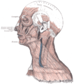 The galea aponeurotica and the temporal fascia
 -
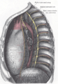
 The fascia of the diaphragm
 -

-


- Visceral Fascia suspends the organs within their cavities and wraps them in layers of connective tissue membranes. Each of the organs is covered in a double layer of fascia; these layers are separated by a thin serous membrane. The outermost wall of the organ is known as the parietal layer, whereas the skin of the organ is known as the visceral layer. The organs have specialized names for their visceral fasciae. In the brain, they are known as meninges; in the heart they are known as pericardia; in the lungs, they are known as pleura; and in the abdomen, they are known as peritonea. [6]
-
 The meninges
 -
 The plurae
 -
 The pericardium and the left cupola of the diaphragm
 -
 The peritoneum and renal fascia


Fascial Dynamics
Fascia is a highly adaptable tissue. Due to its elastic property, superficial fascia can stretch to accomodate the deposition of adipose that accompanies both ordinary and prenatal weight gain. After pregnancy and weight loss, the superficial fascia slowly reverts to its original level of tension.
Deep fascia is less extensible than superficial fascia. It is essentially avascular [7], but is richly innervated with sensory receptors that report the presence of pain (nociceptors); change in movement (proprioceptors); change in pressure and vibration (mechanoreceptors); change in the chemical milieu (chemoreceptors); and fluctuation in temperature (thermoreceptors). [8], [9] Deep fascia is able to respond to sensory input by contracting; by relaxing; or by adding, reducing, or changing its composition through the process of fascial remodeling. [10]
Deep fascia can contract. What happens during the fight-or-flight response is an example of rapid fascial contraction . In response to a real or imagined threat to the organism, the body responds with a temporary increase in the stiffness of the fascia. Bolstered with tensioned fascia, people are able to perform extraordinary feats of strength and speed under emergency conditions. [11] How fascia contracts is still not well understood, but appears to involve the activity of myofibroblasts. Myofibroblasts are fascial cells that are created as a response to mechanical stress. In a two step process, fibroblasts differentiate into proto-myofibroblasts that with continued mechanical stress, become differentiated myofibroblasts. [12] Fibroblasts cannot contract, but myofibroblasts are able to contract in a smooth muscle-like manner. [13] If fascial contraction persists, fibroblasts secrete more collagen into the extracellular matrix, remodeling the fascia to be thicker and less extensible.
The deep fascia can also relax. By monitoring changes in muscular tension, joint position, rate of movement, pressure, and vibration, mechanoreceptors in the deep fascia are capable of initiating relaxation. Deep fascia can relax rapidly in response to sudden muscular overload or rapid movements. Golgi tendon organs operate as a feedback mechanism by causing myofascial relaxation before muscle force becomes so great that tendons might be torn. Pacinian corpuscles monitor the rate of acceleration of movement and will intiate a sudden relaxatory response if movement happens too fast. [14] Deep fascia can also relax slowly as some mechanoreceptors are designed to report changes over a longer period of time. Unlike the Golgi tendon organs, Golgi receptors report joint position independent of muscle contraction. This helps the body to know where the bones are at any given moment. Ruffini endings respond to regular stretching and to slow sustained pressure. In addition to initiating fascial relaxation, they also have been shown to inhibit sympathetic activity by slowing down heart rate and respiration. [15] [16] If fascial relaxation continues, macrophages ingest excess collagen from the extracellular matrix, restoring the fascia to its normal composition and tone.
Visceral fascia is also less extensible than superficial fascia. Due of its suspensory role of the organs, it needs to maintain its tone rather consistently. If it is too lax, it contributes to organ prolapse, yet if it is hypertonic, it restricts proper organ motility. [17]
Fascial Pathology
- Adhesions
- Adhesive capsulitis
- Benign joint hypermobility syndrome
- Calcific tendonitis
- Cardiac tamponade
- Cellulitis
- Compartment syndrome
- Constrictive pericarditis
- Dermatomyositis
- Dupuytren's contracture
- Ehlers-Danlos syndrome
- Eosinophilic fasciitis
- Fibromyalgia
- Hemopneumothorax
- Hemothorax
- Hernia
- Marfan's syndrome
- Meningitis
- Mixed connective tissue disease
- Myofascial pain syndrome
- Necrotizing fasciitis
- Pericardial effusion
- Pericarditis
- Peritonitis
- Plantar fasciitis
- Pleural effusion
- Pleurisy
- Pneumoperitoneum
- Pneumothorax
- Polyarteritis nodosa
- Rheumatoid arthritis
- Scars
- Scleroderma
- Sprain
- Systemic lupus erythematosus
- Tendonitis
- Wegener's granulomatosis
Classification by Region
[18], [19], [20], [21], [22], [23], [24], [25]
References
- ^ Paoletti, Serge (2006). The Fasciae: Anatomy, Dysfunction & Treatment. Seattle, WA: Eastland Press. pp. 151–161. ISBN 0-939616-53-X.
- ^ Skandalakis, John E. (2002). Surgical Anatomy and Technique, 2nd Ed. Atlanta, GA: Springer. pp. 1–2. ISBN 0-38798-752-5.
{{cite book}}: Cite has empty unknown parameter:|1=(help); Unknown parameter|coauthors=ignored (|author=suggested) (help) - ^ Paoletti, Serge (2006). The Fasciae: Anatomy, Dysfunction & Treatment. Seattle, WA: Eastland Press. pp. 23–24. ISBN 0-939616-53-X.
- ^ Hedley, Gil (2005). The Integral Anatomy Series Vol. 1: Skin and Superficial fascia (DVD). Integral Anatomy Productions. Retrieved 2006-07-17.
{{cite AV media}}: Cite has empty unknown parameter:|1=(help) - ^ Hedley, Gil (2005). The Integral Anatomy Series Vol. 2: Deep Fascia and Muscle (DVD). Integral Anatomy Productions. Retrieved 2006-07-17.
{{cite AV media}}: Cite has empty unknown parameter:|1=(help) - ^ Hedley, Gil (2005). The Integral Anatomy Series Vol. 3: Cranial and Visceral Fasciae (DVD). Integral Anatomy Productions. Retrieved 2006-07-17.
{{cite AV media}}: Cite has empty unknown parameter:|1=(help) - ^ Rolf, Ida P. (1989). Rolfing. Rochester, VT: Healing Arts Press. p. 38. ISBN 0-89281-335-0.
- ^ Chaitow, Leon (1988). Soft Tissue Manipulation. Rochester, VT: Healing Arts Press. pp. 26–28. ISBN 0-89281-276-1.
{{cite book}}: Cite has empty unknown parameter:|1=(help) - ^ Schleip, R. (2003). "Fascial plasticity – a new neurobiological explanation: Part 1". Journal of Bodywork and Movement Therapies. 7 (1). Elsevier: 15–19.
{{cite journal}}: Cite has empty unknown parameter:|1=(help) - ^ Myers, Thomas W. (2002). Anatomy Trains. London, UK: Churchill Livingstone. p. 15. ISBN 0-443-06351-6.
- ^ Schleip, R. (2005). "Active fascial contractility: Fascia may be able to contract in a smooth muscle-like manner and thereby influence musculoskeletal dynamics". Medical Hypotheses. 65. Elsevier: 274.
{{cite journal}}: Cite has empty unknown parameter:|1=(help); Unknown parameter|coauthors=ignored (|author=suggested) (help) - ^ Tomasek, J. (2002). "Myofibroblasts and Mechanoregulation of Connective Tissue Remodelling". Molecular Cell Biology. 3. Nature Publishing Group: 350–352.
{{cite journal}}: Cite has empty unknown parameter:|1=(help); Unknown parameter|coauthors=ignored (|author=suggested) (help) - ^ Schleip, R. (2005). "Active fascial contractility: Fascia may be able to contract in a smooth muscle-like manner and thereby influence musculoskeletal dynamics". Medical Hypotheses. 65. Elsevier: 273–277.
{{cite journal}}: Cite has empty unknown parameter:|1=(help); Unknown parameter|coauthors=ignored (|author=suggested) (help) - ^ Chaitow, Leon (1988). Soft Tissue Manipulation. Rochester, VT: Healing Arts Press. pp. 26–27. ISBN 0-89281-276-1.
{{cite book}}: Cite has empty unknown parameter:|1=(help) - ^ Schleip, R. (2003). "Fascial plasticity – a new neurobiological explanation: Part 1". Journal of Bodywork and Movement Therapies. 7 (1). Elsevier: 11–19.
{{cite journal}}: Cite has empty unknown parameter:|1=(help) - ^ Schleip, R. (2003). "Fascial plasticity – a new neurobiological explanation: Part 2". Journal of Bodywork and Movement Therapies. 7 (2). Elsevier: 104–116.
{{cite journal}}: Cite has empty unknown parameter:|1=(help) - ^ Paoletti, Serge (2006). The Fasciae: Anatomy, Dysfunction & Treatment. Seattle, WA: Eastland Press. pp. 146–147. ISBN 0-939616-53-X.
- ^ "Dorlands Medical Dictionary - aponeuroses". Merck Source.
- ^ "Dorlands Medical Dictionary - fasciae". Merck Source.
- ^ "Dorlands Medical Dictionary - ligaments". Merck Source.
- ^ "Dorlands Medical Dictionary - membranes". Merck Source.
- ^ "Dorlands Medical Dictionary - tendons". Merck Source.
- ^ "Fasciae and Aponeuroses - Organized by Region". Department of Anatomy, University of Arkansas for Medical Sciences.
- ^ "Fascia of the Head and Neck". Department of Gross Anatomy at Tufts University.
- ^ "Viscera and Fascia Tables". The University of Michigan - Medical Gross Anatomy.
See also
External links
- Fascia Research
- Fascia Research Congress
- lesson1layersofbody at The Anatomy Lesson by Wesley Norman (Georgetown University)