
Longus colli muscle: Difference between revisions
m cleanup (wikitables, html markup, layout, etc.) |
Oreo Priest (talk | contribs) +translation of name |
||
| Line 17: | Line 17: | ||
| DorlandsSuf = 12549740 |
| DorlandsSuf = 12549740 |
||
}} |
}} |
||
The '''Longus colli muscle''' is a [[muscle]] of the [[human body]]. |
The '''Longus colli muscle''' (Latin for ''long muscle of the neck'') is a [[muscle]] of the [[human body]]. |
||
The Longus colli is situated on the anterior surface of the [[vertebral column]], between the [[atlas (anatomy)|atlas]] and the third [[thoracic vertebra]]. |
The Longus colli is situated on the anterior surface of the [[vertebral column]], between the [[atlas (anatomy)|atlas]] and the third [[thoracic vertebra]]. |
||
Revision as of 19:08, 26 February 2015
| Longus colli muscle | |
|---|---|
 The anterior vertebral muscles. (Longus colli labeled vertically at center left and center right.) | |
| Details | |
| Origin | Transverse processes of C-5 to T-3 |
| Insertion | Anterior arch of the atlas |
| Artery | Ascending Pharyngeal and Vertebral Arteries |
| Nerve | C2-C6 |
| Actions | Flexes the neck and head |
| Identifiers | |
| Latin | musculus longus colli |
| TA98 | A04.2.01.002 |
| TA2 | 2148 |
| FMA | 13370 |
| Anatomical terms of muscle | |
The Longus colli muscle (Latin for long muscle of the neck) is a muscle of the human body.
The Longus colli is situated on the anterior surface of the vertebral column, between the atlas and the third thoracic vertebra.
It is broad in the middle, narrow and pointed at either end, and consists of three portions, a superior oblique, an inferior oblique, and a vertical.
- The superior oblique portion arises from the anterior tubercles of the transverse processes of the third, fourth, and fifth cervical vertebræ and, ascending obliquely with a medial inclination, is inserted by a narrow tendon into the tubercle on the anterior arch of the atlas.
- The inferior oblique portion, the smallest part of the muscle, arises from the front of the bodies of the first two or three thoracic vertebræ; and, ascending obliquely in a lateral direction, is inserted into the anterior tubercles of the transverse processes of the fifth and sixth cervical vertebræ.
- The vertical portion arises, below, from the front of the bodies of the upper three thoracic and lower three cervical vertebræ, and is inserted into the front of the bodies of the second, third, and fourth cervical vertebræ.
Clinical significance
It is commonly injured in rear end whiplash injuries, usually resulting from a car crash.
This muscle is in front of the spine and is thought by some scientists that it may cause some whiplash patients to have an unnatural lack of curvature in the patients' neck.
Additional Images
-
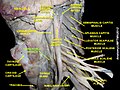
 Longus colli muscle
 -
 Longus colli muscle
 -
 Longus colli muscle


External links
- Template:MuscleLoyola
- . GPnotebook https://www.gpnotebook.co.uk/simplepage.cfm?ID=946536507.
{{cite web}}: Missing or empty|title=(help) - Template:EMedicineDictionary
- PTCentral
- spinal injury database
![]() This article incorporates text in the public domain from page 394 of the 20th edition of Gray's Anatomy (1918)
This article incorporates text in the public domain from page 394 of the 20th edition of Gray's Anatomy (1918)