


Abstract
We studied the crystal structure and spin state of MnP under high pressure with synchrotron x-ray diffraction and x-ray emission spectroscopy (XES). MnP has an exceedingly strong anisotropy in compressibility, with the primary compressible direction along the b axis of the Pnma structure. XES reveals a pressure-driven quenching of the spin state in MnP. First-principles calculations suggest that the strongly anisotropic compression behavior significantly enhances the dispersion of the Mn d-orbitals and the splitting of the d-orbital levels compared to the hypothetical isotropic compression behavior. Thus, we propose spin quenching results mainly from the significant enhancement of the itinerancy of d electrons and partly from spin rearrangement occurring in the split d-orbital levels near the Fermi level. This explains the fast suppression of magnetic ordering in MnP under high pressure. The spin quenching lags behind the occurrence of superconductivity at ∼8 GPa implying that spin fluctuations govern the electron pairing for superconductivity.
Export citation and abstract BibTeX RIS

Original content from this work may be used under the terms of the Creative Commons Attribution 3.0 licence. Any further distribution of this work must maintain attribution to the author(s) and the title of the work, journal citation and DOI.
1. Introduction
The cuprate and the iron-based superconductors possess high Tc [1, 2] and the pairing mechanism is considered closely related to spin fluctuations [3–5]. In their respective phase diagrams, the superconductivity emerges when the long-range magnetic order is suppressed by chemical doping or pressure [3, 6–8]. Spin fluctuations occurring near the disappearance of magnetic order are suggested to play a vital role in mediating Copper pairs. In addition, the organic superconductors and the heavy-fermion superconductors also exhibit similar spin-fluctuation-mediated superconductivity [9–11].
Recently, CrAs and MnP were found to exhibit superconductivity by successfully suppressing their magnetism under high pressure [12, 13]. MnP is regarded as the first discovered manganese-based superconductor. This is a surprising result as inducing superconductivity in manganese-based materials was thought impossible due to their high degree of magnetism. Despite abundant of new efforts made by scientists after the discovery of the superconductivity in MnP, superconductivity in manganese-based materials is still very rare as of today. Besides MnP, only BaMnBi2 was found to be superconducting under pressure [14] while MnAs and MnSb were predicted to be potential superconductors under pressure [15].
The pressure-induced superconductivity in MnP is highly correlated with the magnetic quantum critical point (QCP) as the superconductivity exists only in a narrow pressure window where the magnetic order just vanishes [13]. The coefficient of the T2 term in its resistivity is significantly enhanced near the superconductivity, indicating a strong electronic correlated effect at the QCP and unconventional superconducting nature [13].
To explore the detailed relationship between magnetism and superconductivity in MnP, Matsuda et al and Khasanov et al conducted high-pressure neutron diffraction and muon-spin rotation experiments [16, 17] while Xu et al calculated the magnetic and electronic structures of MnP at different pressures using first-principles methods [18]. From their results, the magnetic ground state of MnP at ambient pressure was identified as a ferromagnetic order below 290 K followed by a helical order below 50 K; both of which were suppressed to lower temperatures under pressure. At 1.2 GPa the helical order was fully suppressed, and at 1.8–2.0 GPa the ferromagnetic order became a conical structure. At 3.8 GPa, a new helical magnetic order below 208 K replaced the conical magnetic order as the magnetic ground state. The magnetic structure above 3.8 GPa was not reported, but it is likely that the occurrence of superconductivity at ∼8 GPa was accompanied by the further suppression of the new helical magnetic order, which is reflected in the continuous suppression of a kink in resistivity [13]. To understand the origin of superconductivity, it is important to understand the magnetic behavior near the occurrence of superconductivity.
Compared with low flux neutrons, synchrotron x-rays have great advantages in terms of the flux and sharpness of beam to probe small-volume samples under extremely high pressure. We employed a diamond anvil cell (DAC) and related synchrotron x-ray techniques, including x-ray diffraction (XRD) and x-ray emission spectroscopy (XES), to study MnP at pressures higher than 3.8 GPa. XES has been widely used to detect the spin state of materials at ambient and high pressures [19, 20], and in this study it helped our fundamental understanding of the magnetism of MnP over a wide pressure range.
Here, we report the crystal structure and spin state of MnP under high pressure which provides a new perspective for understanding the origin of the superconductivity in MnP under high pressure. We argue that the strongly anisotropic compression behavior observed at high pressures brings on spin quenching in MnP and this plays a pivotal role for the suppression of magnetic ordering and occurrence of superconductivity.
2. Experimental section
The powder sample used for this research was synthesized by a solid-state reaction. Elemental Mn and P powders in stoichiometry were mixed, fully ground, and sealed in a silica tube. All procedures were performed in a glove box under an argon atmosphere (both H2O and O2 were limited below 0.1 ppm). The tube containing the elemental mixture was sealed under vacuum at 10−4 mbar and then heated to 500 °C and kept there for 20 h. After the tube was slowly cooled to room temperature, dark gray powder was obtained. A single phase product of MnP was confirmed by XRD with no other phase detected.
The angle-dispersive XRD experiments under high pressure were performed at the HPCAT beamline 16-BM-D at the Advanced Photon Source, Argonne National Laboratory. A MAR345 image plate detector was equipped in the beamline and the diffraction was processed in the transmission mode. The x-ray energy for the experiments was set to 40.443 keV. The application of high pressure was realized with a pair of 300 μm-cutlet diamonds mounted on a symmetric DAC. A T301 stainless steel sheet was used as a gasket and silicon oil was the pressure-transmitting medium. The pressure-transmitting medium was sealed in a 150 μm diameter hole pre-drilled on the gasket for transmitting a quasi-hydrostatic pressure to the MnP sample. The drilling used a laser micro-machining system developed by HPCAT [21]. The pressure was measured with the fluorescence of a ruby, which was put in the same pressure-transmitting medium environment with the sample. The fluorescence peak was sharp at pressures below 30 GPa indicating good hydrostatic character, while at higher pressures it gradually converted and broadened.
The XES experiments under high pressure were carried out at the HPCAT beamline 16-ID-D at the Advanced Photon Source, Argonne National Laboratory. For the transmission of the outgoing x-rays, a beryllium gasket was used and the pressure-transmitting medium was also silicon oil.
3. Results and discussion
MnP adopts an orthorhombic, distorted NiAs structure at ambient pressure. Two-dimensional XRD patterns collected at 0.1 GPa and 40.3 GPa are displayed in figures 1(a) and (b). At 0.1 GPa the diffraction rings are composed of numerous diffraction spots, while at 40.3 GPa the diffraction rings become continuous due to further cracking of crystal grains or pressure gradient. Ideally, homogeneous diffraction rings will guarantee the accuracy of the integrated peak intensities, which is important to the correct determination of the atomic coordinates in the unit cell. In contrast, the diffraction rings composed of spots in our diffraction patterns yielded slight errors on the integrated peak intensities, which results in nonsystematic variety of atomic positions (supporting information is available online at stacks.iop.org/NJP/20/023012/mmedia). The two-dimensional XRD data were reduced to one-dimensional (intensity versus 2θ) using the program DIOPTAS [22]. The obtained one-dimensional XRD patterns for MnP are presented in figure 1(c). The overall diffraction pattern of MnP appears to remain the same but the Bragg peaks shift gradually with increasing pressure, suggesting no phase transition in this pressure range. With more careful analysis, we found that the evolution of the diffraction pattern unveiled a strong anisotropy in compressibility. This is manifested by the fast-shifting Bragg peaks which merge with or split from other peaks upon raising the pressure. In the diffraction patterns, the (00l), (h00) and (h0l) Bragg peaks shift only slightly to higher angles with increasing pressure, but the peaks of k ≠ 0 shift much faster. In figure 1(c), the (111) peak shifts much faster than the (002) and (200) peaks with pressure.

Figure 1. Two-dimensional x-ray diffraction (XRD) patterns collected at (a) 0.1 GPa and (b) 40.3 GPa, and the reduced one-dimensional XRD patterns (c) collected at the pressures from 0.1 to 40.3 GPa. Note that the (002) and (200) peaks shift much slower than the (111) peak with pressure.
Download figure:
Standard image High-resolution imageThe XRD data were refined with the Rietveld method using the software EXPGUI [23], a graphical user interface for GSAS [24]. All the data collected at different pressures can be well fitted with the Pnma structure, the pristine structure at ambient pressure. Thus, the structural stability of MnP under high pressure is proved by these refinements. As a representative, the Rietveld refinement results for 10.2 GPa are shown in figure 2(a). Excellent agreement between the experimental and simulated data is presented by the straight difference line. We also refined the cell parameters and atomic coordinates of MnP at different pressures. The compressions of the a, b, and c axes are plotted in figure 2(b). From this plot, the compression behavior is strongly anisotropic as expected from initial analysis based on the peak shift. At 40 GPa, the cell shrinks along the b axis by nearly 11%, while along the a and c axes by only 2%–3%. With increasing pressure the anisotropic compression behavior with more structural deformation becomes substantial.

Figure 2. (a) Rietveld refinement results for the diffraction data collected at a representative pressure of 10.2 GPa. (b) Compressions along the a, b, and c axes for MnP. Dashed lines are guides for the eyes. (c) Unit cell volume as a function of pressure. The red solid line represents a fit to the third-order Birch–Murnaghan equation of state.
Download figure:
Standard image High-resolution imageWe noticed in a similar work completed by Yu et al, their experimental results are slightly different from ours as they observed a phase transition at ∼15 GPa [25]. We suggest the high pressure phase observed above 15 GPa may be a metastable phase since the ab initio structure prediction supports there is no phase transitions below 55 GPa [25]. The sample stoichiometry may also play a role in the difference of phase transition behavior, since the MnP sample used by Yu et al was grown from Sn flux but the sample we used was made by solid-state reaction. The much larger compressibility of the b-axis is consistent in our and their results.
Compression is often accompanied with a band broadening in the electronic structure of materials, by which the electronic and magnetic properties can be tuned, for instance, by narrowing the band gap or metallization [26–28]. For MnP, the strongly anisotropic compression behavior brings drastic changes in the electronic structure and gives rise to its electronic phase diagram over the pressure. The unit cell volume as a function of pressure for MnP, is shown in figure 2(c). The data can be fitted (the red solid line in figure 2(c)) with the third-order Birch–Murnaghan equation of state [29]
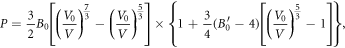
where B0 is the bulk modulus,  the derivative of the bulk modulus at ambient pressure, and V0 is the volume at ambient pressure. With V0 fixed as 98.85 Å3, we obtained B0 = 116 ± 12 GPa and
  = 4.2 ± 0.8 GPa. The fact that all the data can be fitted with a single curve indicates no pressure-induced isostructural volume collapse in MnP.
The crystal structure of MnP at ambient pressure consists of a three-dimensional network of MnP6 octahedra, figure 3(a). Since each Mn and P atom occupy only one independent crystallographic site, all MnP6 octahedra in the structure are equivalent. The coordination environment of Mn is shown in figure 3(b). The analysis of the pressure effect on the crystal structure can be simplified by analysis of the MnP6 octahedra. The primary compressible direction for MnP is marked in figure 3(b). It should be noted that the MnP6 octahedron presented in figure 3(b) is already slightly distorted before the application of pressure. For better illustration of the pressure effect on the structure, the P atoms in the MnP6 octahedron are labeled with P(1), P(2), P(3), P(4), P(5), and P(6) in figure 3(b). The Mn atom is located at the center of the MnP6 octahedron hidden behind P(5). The P(m)–Mn–P(n) (m, n = 1, 2, 3, 4, 5, 6) bonding angles are shown as a function of pressure in figure 3(c). In an ideal octahedron, the angle of two neighboring vertices to the body center should be 90°. With the application of pressure the divergence of P(1)–Mn–P(2), P(1)–Mn–P(3), P(2)–Mn–P(4) and P(3)–Mn–P(4) bonding angles from 90° gradually increases while the other bonding angles remain nearly constant. All the Mn–P(n) (n = 1, 2, 3, 4, 5, 6) bond lengths are shortened with pressure but the degree of this effect is very little compared to the anisotropic distortion of the MnP6 octahedra. This indicates that the main contribution of the strong anisotropic compression behavior of the MnP structure is the anisotropic distortion of MnP6 octahedra with increasing pressure.

Figure 3. (a) Crystal structure of MnP at ambient pressure. (b) The evolution of the MnP6 octahedron in MnP before and after pressure applied. The open arrow signs indicate the primary compressible direction at high pressure. (c) P(m)–Mn–P(n) (m, n = 1, 2, 3, 4, 5, 6) bonding angles as a function of pressure.
Download figure:
Standard image High-resolution imageThe Mn Kβ emission spectra, which arise from the 3p → 1s transition, were obtained as a function of pressure up to 40 GPa, as shown in figure 4(a). We observed a main peak (Kβ1,3) around 6489 eV and a satellite peak (Kβ') around 6476 eV. The Kβ emission spectrum of Mn2O3 taken from the [30] is also shown for comparison; where the Mn3+ is in a high spin state for the 3d4 electron configuration (the detailed configuration of unpaired 3d electrons is  and the spin state is S = 2). The satellite Kβ' was suggested to originate from the exchange interaction between the 3d electrons and the 3p core hole left in the final state of the emission process [31]. For the current system, the Kβ' region derives from the final state with antiparallel net spins between the 3p5 hole and the 3d4 valence shell. When one 3d electron pairs with another, on the whole, their spins will neutralize, and thus the total spins in the 3d4 valence shell will reduce. In this way the exchange interaction between the 3p5 hole and the 3d4 valence shell will weaken, which results in a less intense peak of Kβ'.

Figure 4. (a) X-ray emission spectra collected at different pressures up to 40 GPa for MnP. The insets show expanded views of the Kβ1,3 peak and the satellite Kβ' peak. (b) The spin values as a function of pressure calculated according to the integrated relative difference (IRD) method.
Download figure:
Standard image High-resolution imageIn previous experiments, the intensity of Kβ' roughly correlated with the number of unpaired 3d electrons on the transition metal [32]. At ambient pressure, the intensity of Kβ' for MnP is lower than that for Mn2O3, indicating the unpaired 3d electrons in MnP are fewer than in Mn2O3. This is consistent with the striking contrast in the strength of the magnetic moment of Mn for MnP and Mn2O3. Neutron diffraction determined the moment of Mn3+ in MnP as ∼1.3 μB/Mn while in Mn2O3 it was ∼3.8 μB/Mn [33-35], from which we can conclude that the Mn3+ in MnP is in the low spin state for the 3d4 electron configuration,  and S = 1. From the expanded view of the satellite peak Kβ' in the inset of figure 4(a), we see the application of pressure gradually suppresses the intensity of the satellite peak Kβ' of MnP. Following the integrated relative difference method described by Zhu Mao et al [36], we derived the spin quantum number as a function of pressure, as shown in figure 4(b). This plot shows the spin quantum number S decreases rapidly from 0 to ∼16 GPa, and at higher pressures, it slackens its steps and gradually vanishes to zero. At ∼8 GPa (the pressure where superconductivity occurs [13]) the spin is in an intermediate state of S ≈ 0.6. The spin weakening from S = 1 to S ≈ 0.6 brings on a weakening of the magnetic-coupling strength, which vitally induces the suppression of the magnetic ordering in MnP. Moreover, the energy difference between the main peak Kβ1,3 and the satellite peak Kβ' is also related to the 3p–3d exchange interaction [37], therefore, the peak shift of Kβ1,3 can also reflect the spin state transition qualitatively. A clear pressure-induced peak shift for the Kβ1,3 of MnP is visible in the expanded view of the main peak Kβ1,3 in the right inset of figure 4(a).
4. Band structure calculations
To understand the spin quenching induced by high pressure in MnP, we also studied the MnP electronic structure. The band structures of MnP were calculated using a LDA potential without spin-polarization based on the crystal structures at ambient and high pressures. For a comparison, we assumed an isotropic compression behavior for which the unit cell volume and atomic coordinates are equal to the experimental anisotropic compression behavior. From the band structures in figure 5, the d-orbitals of the Mn atoms and the p-orbitals of the P atoms are highly hybridized. With no matter isotropic or anisotropic compression behavior, the dispersion of the Mn d-orbitals becomes larger, which can result in an enhancement of the itinerancy of d electrons and thus, the magnetism is reduced naturally. Meanwhile, the expanding dispersion of the Mn d-orbitals also results in a reduction of the density of states around the Fermi level, which means a reduction of the carrier concentration under high pressure. However, in a previous experiment, the room-temperature resistivity of MnP barely changed with pressure [13]. This result reflects an enhancement of the Fermi velocity under high pressure, since the carrier concentration is reduced but the resistivity does not change. The enhancement of the Fermi velocity further proves the enhancement of the itinerancy of d electrons. The comparison of figures 5(b) and (c) shows that the strongly anisotropic compression behavior can lead to a much more remarkable widening effect on the dispersion of the Mn d-orbitals. Therefore, we suggest the strongly anisotropic compression behavior significantly accelerates the disappearance of magnetism compared with the hypothetical isotropic compression behavior.

Figure 5. Band structures of MnP calculated by the LDA method based on the crystal structures (a) at ambient pressure, (b) at 40 GPa for an hypothetical isotropic compression behavior, and (c) at 40 GPa for the experimentally observed anisotropic compression behavior, respectively. The weight of the Mn–3d state is proportional to the width of the curves.
Download figure:
Standard image High-resolution imageFrom the band structures, we conclude that the splitting of the d-orbital levels around the Fermi level is enhanced when pressure is applied, which may explain the spin quenching from another perspective. Compared with the hypothetical isotropic compression behavior, the strongly anisotropic compression behavior widens the d-orbital splitting more significantly. The more significant widening of the d-orbital splitting induces larger energy gaps between any two of the energy levels of the t2g orbitals. This may lead to a complete spin pairing in the t2g orbitals. A schematic representation of the possible spin rearrangement is shown in figure 6. In fact, the gap width of the d-orbital splitting is thought to be associated with the degree of the geometry distortion of the MX6 octahedron, which has been successfully used to explain the Jahn–Teller distortion [38]. Generally, the more distorted the MX6 octahedron is, the larger the splitting becomes, which is a phenomenological description for the crystal field. Thus, the above analysis on bonding angles consistently leads to the conclusion of spin quenching.

Figure 6. Schematic representation of the d-orbital splitting and the electron configuration for an ideal-octahedron ligand field of low-spin 3d4 cations, and the distorted MnP6 octahedron in MnP at ambient pressure (AP) and high pressure (HP) (from left to right).
Download figure:
Standard image High-resolution image
5. Concluding remarks
MnP is unique as the first manganese-based superconductor, and it is therefore important to understand the relationship between superconductivity and magnetism. Our study demonstrates that the suppression of magnetic ordering in MnP is due to the spin weakening induced by high pressure. The spin state of MnP changes from S = 1 to S = 0 gradually, resulting in a decrease of the Mn magnetic moment and weakening of the magnetic-coupling strength. When the spin adopts an intermediate state and the magnetic coupling is weak enough but not vanished, the spins begin to fluctuate in the lattice, which is thought to mediate the electron pairing of superconductivity. More importantly, a qualitative analysis based on the electronic structures indicates that the spin weakening and quenching is assisted by the strongly anisotropic compression behavior in this compound. The strong anisotropy of the compression behavior further enhances the effect of high pressure on the itinerancy of the d electrons and the widening effect on the d-orbital splitting, which greatly accelerates the spin disappearance. This explains why magnetism in MnP is not as robust as in other manganese-based systems under high pressure and why superconductivity can arise in MnP.
Acknowledgments
This work was financially supported by the National Nature Science Foundation of China under Contract Nos. U1530402, 11374137, and 11525417. WY and MHK also acknowledge financial support from the US Department of Energy, Office of Basic Energy Sciences, x-ray Scattering Core Program under Contract No. DE-FG02-99ER45775. HPCAT operations are supported by the US Department of Energy, National Nuclear Security Administration under Contract No. DE-NA0001974 and the US Department of Energy, Office of Basic Energy Science under Contract No. DE-FG02-99ER45775, with partial instrumentation funding from National Science Foundation. The Advanced Photon Source is supported by the US Department of Energy, Office of Basic Energy Sciences under Contract No. DE-AC02-06CH11357. The sample preparation conducted at the Materials Science Division, Argonne National Laboratory is supported by the US Department of Energy, Office of Science, Materials Sciences and Engineering.