SlideShare a Scribd company logo
ST. IGNATIUS INSTITUTE OF HEALTH SCIENCES, HONAVAR
Mr. Shivalinga Talekar
Lecture
Dept. Medical Surgical Nursing
St. Ignatius institute of health sciences, Honavar.
CRANIAL NERVE AND THE EXAMINATION
Sl.
No
Cranial Nerve The Examination
I. OLFACTORY NERVE Not routinely tested unless the patient complains
of a loss of sense of smell (anosmia)
“Have you noticed any recent change in your
sense of smell?”
Casual: take a nearby odorous object (e.g. coffee
or chocolate) and ask the patient if it smells
normal.
Formal: a series of identical bottles containing
recognizable smells are used. The patient is asked
to identify them.
Commonly used agents: coffee, vanilla, vinegar.
Test each nostril separately and determine if any
loss of smell is unilateral or bilateral.
II. OPTIC NERVE VISUAL ACUITY, VISUAL FIELDS & THE
PUPILS.
 Sit directly facing the patient, approximately one
meter away.
 Ask the patient to cover their left eye with their
left hand.
You should cover your right eye and be staring
directly at the patient
 Ask the patient to look into your eye and not
move their head or eyes.
 Ask the patient to tell you when they can see your
fingertip moving.
 Position your fingertip at the outer border of one
of the quadrants of your visual field.
 Slowly bring your fingertip inwards, towards the
center of your visual field until the patient sees it.
 Repeat this process for each quadrant – at
10 o’clock /2 o’clock / 4 o’clock / 8 o’clock.
 Repeat the same assessment process on the other
eye.
ST. IGNATIUS INSTITUTE OF HEALTH SCIENCES, HONAVAR
Mr. Shivalinga Talekar
Lecture
Dept. Medical Surgical Nursing
St. Ignatius institute of health sciences, Honavar.
III,
IV
&
VI
OCULOMOTOR(III),
TROCHLEAR(IV) and
ABDUCENS(VI)
Examining Eye Movements
 Hold your finger about 30cm directly in front of
the patient’s eyes and ask them to look at it. Look
at the eyes in the primary position for any
deviation or abnormal movements.
 Ask the patient to keep their head still and follow
your finger with their eyes.
 Ask the patient to report any double vision.
 Move your finger through the various axes of
eye movement (“H” shape).
 Observe for restriction of eye movement
 and note any nystagmus.
V TRIGEMINAL Inspection
Inspect the patient’s face—wasting of the
temporalis will show as hollowing above the
zygomatic arch.
Testing motor function
• Ask the patient to clench their teeth and feel both
sides for the bulge
of the masseter and temporalis.
• Ask the patient to open their mouth wide—the jaw
will deviate
towards the side of a V lesion.
• Again ask them to open their mouth but provide
resistance
by holding their jaw closed with one of your hands.
Testing sensory function
• Distribution of the sensory branches of the
trigeminal nerve.
V1 = ophthalmic, V2 = maxillary, V3 = mandibular.
Assess light touch for each branch and ask the
patient to say ‘yes’ if
they can feel it.
• Choose three spots to test on each side to make the
examination
easy to remember—forehead, cheek, and mid-way
along jaw.
• For each branch, compare left to right.
• Test pin-prick sensation at the same spots using a
sterile pin.
• Temperature sensation is not routinely tested
consider only if
abnormalities in light touch or pin-prick are found.
Use specimen
tubes or other small containers full of warm or cold
water.
Jaw jerk and corneal reflex
ST. IGNATIUS INSTITUTE OF HEALTH SCIENCES, HONAVAR
Mr. Shivalinga Talekar
Lecture
Dept. Medical Surgical Nursing
St. Ignatius institute of health sciences, Honavar.
VII FACIAL Muscles of facial expression
• Look at the patient’s face at rest. Look for
asymmetry in the nasolabial folds, angles of the
mouth, and forehead wrinkles.
• Ask the patient to raise their eyebrows (‘look up!’)
and watch the forehead wrinkle.
• Attempt to press their eyebrows down and note
any weakness.
• Ask the patient to ‘close your eyes tightly’. Watch,
then test against resistance with your finger and
thumb. ‘Don’t let me pull them apart.’
• Ask the patient to blow out their cheeks. Watch for
air escaping on one side.
• Ask the patient to bare their teeth. ‘Show me your
teeth!’ Look for asymmetry.
• Ask the patient to purse their lips. ‘Whistle for
me!’ Look for asymmetry.
ST. IGNATIUS INSTITUTE OF HEALTH SCIENCES, HONAVAR
Mr. Shivalinga Talekar
Lecture
Dept. Medical Surgical Nursing
St. Ignatius institute of health sciences, Honavar.
VIII VESTIBULOCOCHLEA Enquire first about symptoms—hearing
loss/changes or balance problems.
Begin by inspecting each ear.
Hearing
Test each ear separately. Cover one by pressing on
the tragus. Simple test of hearing
• Whisper a number into one ear and ask the patient
to repeat it.
• Repeat with the other ear.
• Be careful to whisper at the same volume in each
ear and at the same distance (about 60cm).
Rinne’s Test
• Tap a tuning fork and hold adjacent to the ear (air
conduction).
• Then apply the base of the tuning fork to the
mastoid process (bone conduction).
• Ask the patient which position sounds louder.
• Normal = air conduction > bone conduction =
‘Rinne’s positive’
• In neural deafness, Rinne’s test will remain
positive
• In conductive deafness, the findings are reversed
(bone > air).
Weber’s test
• Tap a tuning fork and hold the base against the
vertex or forehead at the midline.
• Ask the patient if it sounds louder on one side.
• In neural deafness, the tone is heard better in the
intact ear
• In conductive deafness, the tone is heard better in
the affected ear.
IX
and
X
GLOSSOPHARYNGEAL and
VAGUS NERVE
Pharynx
• Ask the patient to open their mouth and inspect
the uvula (use a tongue depressor if necessary). Is it
central or deviated to one side?
• Ask the patient to say ‘aah’. Watch the uvula. It
should move upwards centrally. Does it deviate to
one side?
Gag reflex
This is unpleasant for the patient and should only be
tested if a IX or X nerve lesion is suspected
(afferent signal = IX, efferent = X).
• With the patient’s mouth open wide, gently touch
the posterior pharyngeal wall on one side with a
tongue depressor.
• Watch the uvula (it should lift up).
ST. IGNATIUS INSTITUTE OF HEALTH SCIENCES, HONAVAR
Mr. Shivalinga Talekar
Lecture
Dept. Medical Surgical Nursing
St. Ignatius institute of health sciences, Honavar.
• Repeat on the opposite side.
XI ACCESSORY Applied anatomy
• Sensory: none
• Motor: sternocleidomastoids and upper part of
trapezii
• Note that each cerebral hemisphere controls the
ipsilateral sternocleidomastoid and the
contralateral trapezius.
Inspect the sternocleidomastoids.
Look for wasting, fasciculation, hypertrophy, and
any abnormal head position.
• Ask the patient to raise their shoulders and
observe.
• Ask the patient to raise again, using your hands on
their shoulders to provide resistance.
• Ask the patient to turn their head to each side, first
without and then with resistance (use your hand on
their cheek).
 Be sure to press against the patient’s cheek.
Lateral pressure to the jaw can cause pain and
injury.
XII HYPOGLOSSAL Applied anatomy
Sensory: none
Motor: muscles of the tongue
Examination
• Ask the patient to open wide and inspect the
tongue on the floor of the mouth.
Look for size and evidence of fasciculation.
• Ask the patient to protrude the tongue. Look for
deviation or
abnormal movements.
• Ask the patient to move the tongue in and out
repeatedly, then side to side.
• To test for weakness, place your finger on the
patient’s cheek
and ask them to push against it from the inside
using their tongue.

More Related Content

Anatomy and physiology of CRANIAL NERVE AND THE EXAMINATION.pdf

  • 1. ST. IGNATIUS INSTITUTE OF HEALTH SCIENCES, HONAVAR Mr. Shivalinga Talekar Lecture Dept. Medical Surgical Nursing St. Ignatius institute of health sciences, Honavar. CRANIAL NERVE AND THE EXAMINATION Sl. No Cranial Nerve The Examination I. OLFACTORY NERVE Not routinely tested unless the patient complains of a loss of sense of smell (anosmia) “Have you noticed any recent change in your sense of smell?” Casual: take a nearby odorous object (e.g. coffee or chocolate) and ask the patient if it smells normal. Formal: a series of identical bottles containing recognizable smells are used. The patient is asked to identify them. Commonly used agents: coffee, vanilla, vinegar. Test each nostril separately and determine if any loss of smell is unilateral or bilateral. II. OPTIC NERVE VISUAL ACUITY, VISUAL FIELDS & THE PUPILS.  Sit directly facing the patient, approximately one meter away.  Ask the patient to cover their left eye with their left hand. You should cover your right eye and be staring directly at the patient  Ask the patient to look into your eye and not move their head or eyes.  Ask the patient to tell you when they can see your fingertip moving.  Position your fingertip at the outer border of one of the quadrants of your visual field.  Slowly bring your fingertip inwards, towards the center of your visual field until the patient sees it.  Repeat this process for each quadrant – at 10 o’clock /2 o’clock / 4 o’clock / 8 o’clock.  Repeat the same assessment process on the other eye.
  • 2. ST. IGNATIUS INSTITUTE OF HEALTH SCIENCES, HONAVAR Mr. Shivalinga Talekar Lecture Dept. Medical Surgical Nursing St. Ignatius institute of health sciences, Honavar. III, IV & VI OCULOMOTOR(III), TROCHLEAR(IV) and ABDUCENS(VI) Examining Eye Movements  Hold your finger about 30cm directly in front of the patient’s eyes and ask them to look at it. Look at the eyes in the primary position for any deviation or abnormal movements.  Ask the patient to keep their head still and follow your finger with their eyes.  Ask the patient to report any double vision.  Move your finger through the various axes of eye movement (“H” shape).  Observe for restriction of eye movement  and note any nystagmus. V TRIGEMINAL Inspection Inspect the patient’s face—wasting of the temporalis will show as hollowing above the zygomatic arch. Testing motor function • Ask the patient to clench their teeth and feel both sides for the bulge of the masseter and temporalis. • Ask the patient to open their mouth wide—the jaw will deviate towards the side of a V lesion. • Again ask them to open their mouth but provide resistance by holding their jaw closed with one of your hands. Testing sensory function • Distribution of the sensory branches of the trigeminal nerve. V1 = ophthalmic, V2 = maxillary, V3 = mandibular. Assess light touch for each branch and ask the patient to say ‘yes’ if they can feel it. • Choose three spots to test on each side to make the examination easy to remember—forehead, cheek, and mid-way along jaw. • For each branch, compare left to right. • Test pin-prick sensation at the same spots using a sterile pin. • Temperature sensation is not routinely tested consider only if abnormalities in light touch or pin-prick are found. Use specimen tubes or other small containers full of warm or cold water. Jaw jerk and corneal reflex
  • 3. ST. IGNATIUS INSTITUTE OF HEALTH SCIENCES, HONAVAR Mr. Shivalinga Talekar Lecture Dept. Medical Surgical Nursing St. Ignatius institute of health sciences, Honavar. VII FACIAL Muscles of facial expression • Look at the patient’s face at rest. Look for asymmetry in the nasolabial folds, angles of the mouth, and forehead wrinkles. • Ask the patient to raise their eyebrows (‘look up!’) and watch the forehead wrinkle. • Attempt to press their eyebrows down and note any weakness. • Ask the patient to ‘close your eyes tightly’. Watch, then test against resistance with your finger and thumb. ‘Don’t let me pull them apart.’ • Ask the patient to blow out their cheeks. Watch for air escaping on one side. • Ask the patient to bare their teeth. ‘Show me your teeth!’ Look for asymmetry. • Ask the patient to purse their lips. ‘Whistle for me!’ Look for asymmetry.
  • 4. ST. IGNATIUS INSTITUTE OF HEALTH SCIENCES, HONAVAR Mr. Shivalinga Talekar Lecture Dept. Medical Surgical Nursing St. Ignatius institute of health sciences, Honavar. VIII VESTIBULOCOCHLEA Enquire first about symptoms—hearing loss/changes or balance problems. Begin by inspecting each ear. Hearing Test each ear separately. Cover one by pressing on the tragus. Simple test of hearing • Whisper a number into one ear and ask the patient to repeat it. • Repeat with the other ear. • Be careful to whisper at the same volume in each ear and at the same distance (about 60cm). Rinne’s Test • Tap a tuning fork and hold adjacent to the ear (air conduction). • Then apply the base of the tuning fork to the mastoid process (bone conduction). • Ask the patient which position sounds louder. • Normal = air conduction > bone conduction = ‘Rinne’s positive’ • In neural deafness, Rinne’s test will remain positive • In conductive deafness, the findings are reversed (bone > air). Weber’s test • Tap a tuning fork and hold the base against the vertex or forehead at the midline. • Ask the patient if it sounds louder on one side. • In neural deafness, the tone is heard better in the intact ear • In conductive deafness, the tone is heard better in the affected ear. IX and X GLOSSOPHARYNGEAL and VAGUS NERVE Pharynx • Ask the patient to open their mouth and inspect the uvula (use a tongue depressor if necessary). Is it central or deviated to one side? • Ask the patient to say ‘aah’. Watch the uvula. It should move upwards centrally. Does it deviate to one side? Gag reflex This is unpleasant for the patient and should only be tested if a IX or X nerve lesion is suspected (afferent signal = IX, efferent = X). • With the patient’s mouth open wide, gently touch the posterior pharyngeal wall on one side with a tongue depressor. • Watch the uvula (it should lift up).
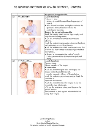
  • 5. ST. IGNATIUS INSTITUTE OF HEALTH SCIENCES, HONAVAR Mr. Shivalinga Talekar Lecture Dept. Medical Surgical Nursing St. Ignatius institute of health sciences, Honavar. • Repeat on the opposite side. XI ACCESSORY Applied anatomy • Sensory: none • Motor: sternocleidomastoids and upper part of trapezii • Note that each cerebral hemisphere controls the ipsilateral sternocleidomastoid and the contralateral trapezius. Inspect the sternocleidomastoids. Look for wasting, fasciculation, hypertrophy, and any abnormal head position. • Ask the patient to raise their shoulders and observe. • Ask the patient to raise again, using your hands on their shoulders to provide resistance. • Ask the patient to turn their head to each side, first without and then with resistance (use your hand on their cheek).  Be sure to press against the patient’s cheek. Lateral pressure to the jaw can cause pain and injury. XII HYPOGLOSSAL Applied anatomy Sensory: none Motor: muscles of the tongue Examination • Ask the patient to open wide and inspect the tongue on the floor of the mouth. Look for size and evidence of fasciculation. • Ask the patient to protrude the tongue. Look for deviation or abnormal movements. • Ask the patient to move the tongue in and out repeatedly, then side to side. • To test for weakness, place your finger on the patient’s cheek and ask them to push against it from the inside using their tongue.